Upper And Lower Extremity Fractures
Article Sections
- Upper extremity anatomy
- Lower extremity anatomy
- General fracture evaluation and management
- Upper extremity fractures
- Clavicle fracture
- Humerus fractures
- Elbow/forearm fractures
- Lower extremity fractures
- Hip fractures
- Midshaft femur fractures
- Tibia fractures
- Ankle fracture (vs ankle sprain)
- Jones fracture (proximal fifth metatarsal fracture)
- Calcaneal fracture
- Stress fracture
- Complications
- Summary
Fractures of the upper and lower extremities vary by mechanism, associated neurovascular risks, and management requirements. This article reviews the anatomy of the upper and lower extremities, general principles of fracture assessment and management, and the unique features of different fracture types.
Upper extremity anatomy
Axillary, brachial, radial, and ulnar arteries (Figure 1).
Major nerves- Brachial plexus (Figure 2): A network of nerves originating from the spinal cord (C5-T1) that provides motor and sensory innervation to the shoulder, arm, and hand. Divides into 5 major peripheral nerves: the axillary, musculocutaneous, radial, median, and ulnar nerves.
- Axillary nerve (Figure 3): Innervates the deltoid and teres minor muscles, allowing shoulder abduction and external rotation. Provides sensation to the skin over the lateral shoulder (
Continue Learning with UWorld
Get the full Upper And Lower Extremity Fractures article plus rich visuals, real-world cases, and in-depth insights from medical experts, all available through the UWorld Medical Library.
Unlock Full AccessFigures
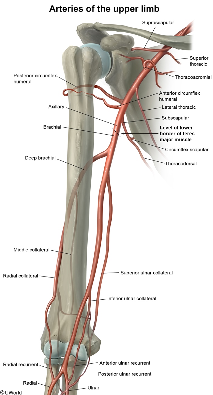
Figure 1
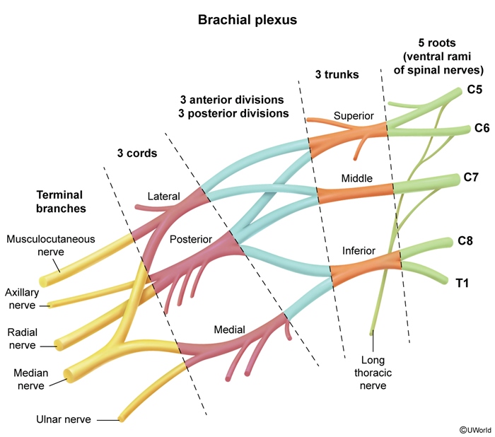
Figure 2
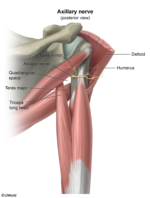
Figure 3
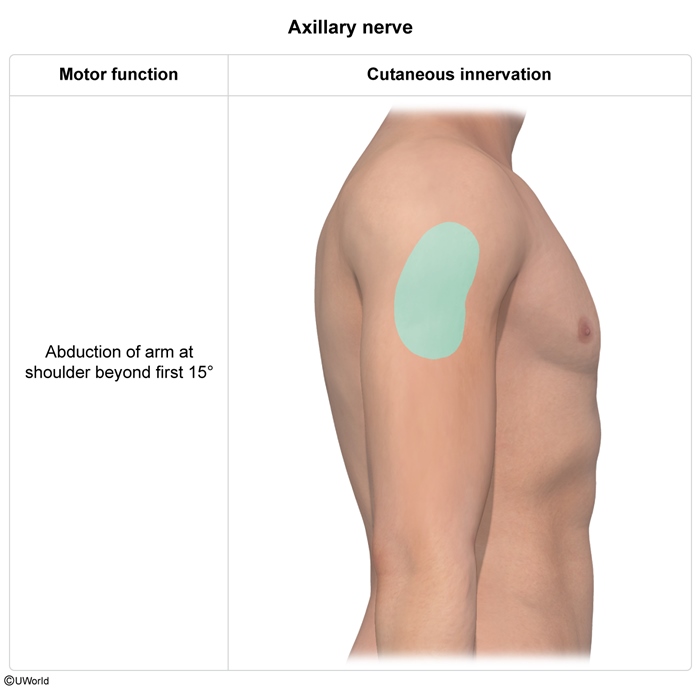
Figure 4
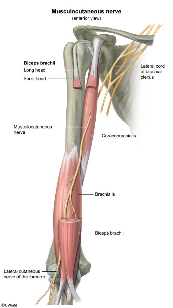
Figure 5
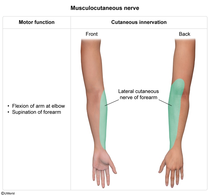
Figure 6
Medical Image
Medical Illustration