Hip Dislocations
Article Sections
Introduction
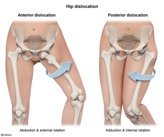
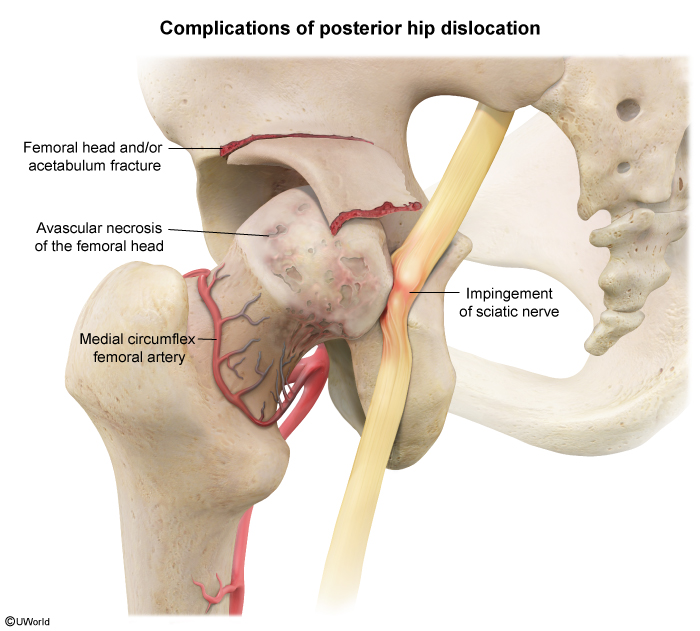
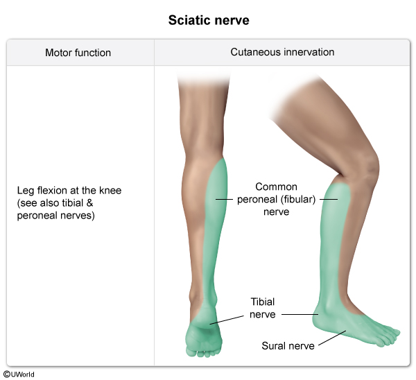
Hip dislocations occur when the femoral head is displaced from the acetabulum (hip socket), often due to a high energy trauma. It is a medical emergency that requires prompt intervention (ie, reduction) due to the risk of neurovascular compromise, which can lead to avascular necrosis of the femoral head and nerve palsies.
Anatomy
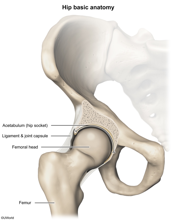
The hip, a stable ball-and-socket joint formed by the femoral head (the ball) and the acetabulum (the socket), is able to withstand substantial mechanical forces (Figure 1). The joint's stability is supported by the depth of the acetabulum, the cartilaginous labrum, the fibrous joint capsule, and the surrounding muscles (eg, gluteal muscles, rectus femoris) and ligaments (eg, iliofemoral, ischiofemoral).
The dominant blood supply to the femoral head and neck is from the medial femoral circumflex artery and its branches (eg, retinacular arteries). Injury to these vessels from a hip fracture or dislocation can impair perfusion and result in avascular necrosis (ie, bone ischemia leading to bone death) of the femoral head.
Continue Learning with UWorld
Get the full Hip Dislocations article plus rich visuals, real-world cases, and in-depth insights from medical experts, all available through the UWorld Medical Library.
Unlock Full AccessFigures