Knee Trauma
Article Sections
Introduction
The knee joint, a complex hinge structure, is susceptible to various injuries due to its weight-bearing function and the numerous ligaments, tendons, cartilage, and bones involved in its stability and movement. Traumatic injury to any of these components can significantly impact knee mobility and function.
Overuse injuries of the knee are discussed in more detail in a separate dedicated article.
Anatomy
- Femur: The distal end of the femur has 2 rounded protrusions, the medial and lateral condyles (Figure 1), which articulate with the tibia.
- Tibia: The flat upper surface that interacts with the femoral condyles is the tibial plateau. The prominent area on the anterior surface where the patellar tendon attaches is the tibial tuberosity.
- Patella: This is the large sesamoid bone that facilitates knee extension, protects the joint from direct injury, and improves nourishment of the distal femur articular cartilage.
Continue Learning with UWorld
Get the full Knee Trauma article plus rich visuals, real-world cases, and in-depth insights from medical experts, all available through the UWorld Medical Library.
Unlock Full AccessFigures
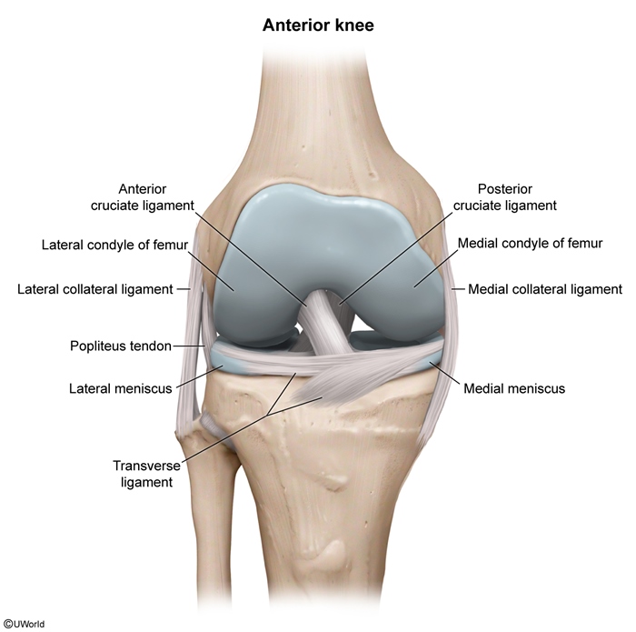
Figure 1
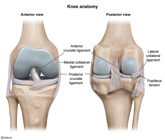
Figure 2
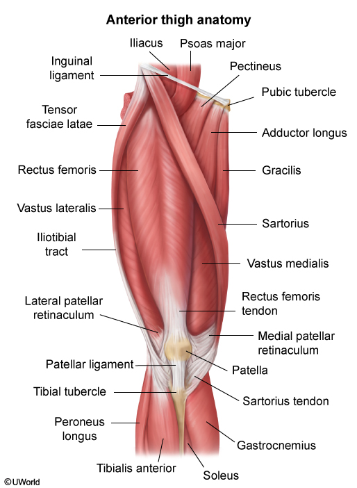
Figure 3
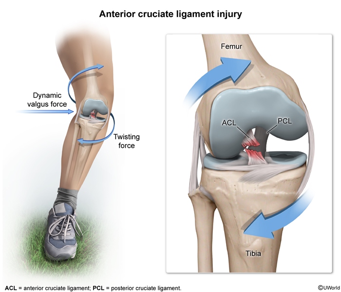
Figure 4
Images
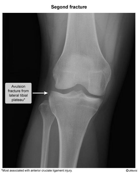
Image 1
Tables
Table 1
Medical Image
Medical Illustration