Atelectasis
Article Sections
Introduction
Atelectasis (Greek: ateles = incomplete, ektasis = expansion) refers to loss of alveolar aeration with corresponding collapse of lung tissue. It appears as a region of opacification on chest imaging, which can range from minimal impairment to significant hypoxemia, pneumonia, and other complications if not resolved.
Obstructive atelectasis
Obstructive (resorptive) atelectasis is the most common form.
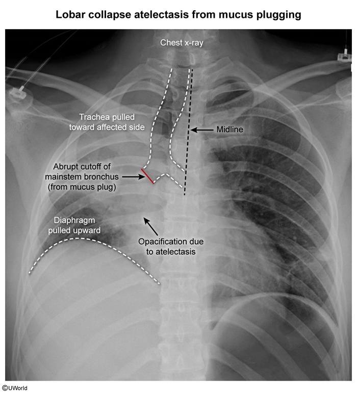
PathogenesisObstructive (resorptive) atelectasis occurs when a bronchus becomes blocked, resulting in loss of ventilation to the downstream lobe. Because the lobe continues to be perfused, alveolar gas is reabsorbed into the bloodstream. As the alveoli become airless, they collapse and appear opaque on chest x-ray (Image 1). This form of atelectasis is associated with:
- Mucus plugging (eg, bronchial infections, pneumonia).
- Aspirated foreign body.
- Tumors or mass lesions (eg, lung cancer).
Continue Learning with UWorld
Get the full Atelectasis article plus rich visuals, real-world cases, and in-depth insights from medical experts, all available through the UWorld Medical Library.
Unlock Full AccessFigures
Images