Congenital Diaphragmatic Hernia
Article Sections
Introduction
Congenital diaphragmatic hernia (CDH) is a rare but life-threatening condition characterized by a defect in the diaphragm through which abdominal viscera herniates into the thorax, compromising pulmonary development and function.
Pathophysiology
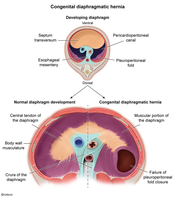
During embryonic development, the diaphragm forms from 4 distinct tissues (Figure 1): the septum transversum, esophageal mesentery, body wall musculature, and pleuroperitoneal folds. The pleuroperitoneal folds extend over and fuse with the other structures to divide the pericardioperitoneal canal into the thoracic and peritoneal cavities.
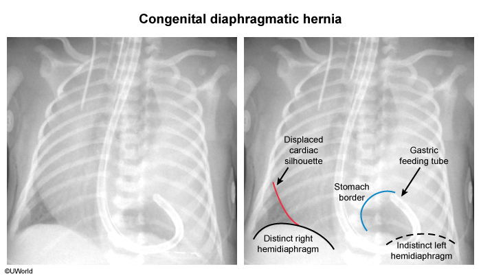
Incomplete fusion results in an abnormal connection between the thorax and the abdomen, allowing bowel, stomach, spleen, and/or liver to herniate into the left thorax. This connection is located on the left in 80%-85% of cases, on the right in 10%-15% of cases, and bilaterally in <2% of cases. When located on the left, the herniated abdominal viscera displace the heart into the right thorax. In addition, the herniated abdominal viscera compress the developing lungs, leading to pulmonary hypoplasia and each of the following:
Continue Learning with UWorld
Get the full Congenital Diaphragmatic Hernia article plus rich visuals, real-world cases, and in-depth insights from medical experts, all available through the UWorld Medical Library.
Unlock Full AccessFigures
Images