Retinal Detachment
Article Sections
Introduction
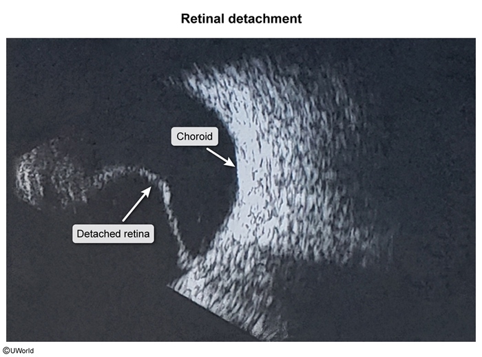
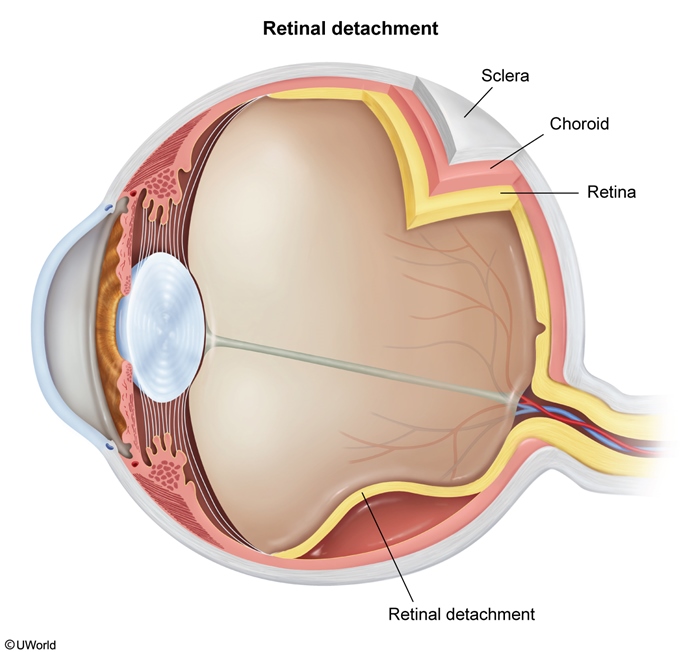
Retinal detachment is characterized by separation of the neurosensory retina (photoreceptors) from the underlying retinal pigment epithelium and choroid that nourish it. Most cases are preceded by a posterior vitreous detachment, which exerts traction on the retina, causing retinal tears and eventual detachment. Patients classically have acute, painless, monocular visual disturbance accompanied by flashes of light and/or floaters.
Anatomy and physiology
The retina is made up of several cellular layers, including the internal limiting membrane, which is the innermost layer located just posterior to the vitreous cavity, followed posteriorly by the ganglion cells, bipolar cells, and photoreceptors. The retinal pigment epithelium is the outermost layer and sits against the choroid. The choroid provides oxygen and nutrition for the photoreceptor layer.
The neurosensory retina (photoreceptors) is responsible for processing light information and transmitting signals through the optic nerve to the brain. The macula, located in the center of the retina, has the highest density of cones and provides sharp, central vision. The peripheral retina has a higher proportion of rods and helps provide vision in dark conditions and peripheral vision.
Continue Learning with UWorld
Get the full Retinal Detachment article plus rich visuals, real-world cases, and in-depth insights from medical experts, all available through the UWorld Medical Library.
Unlock Full AccessFigures
Images