Cauda Equina Syndrome
Article Sections
Introduction
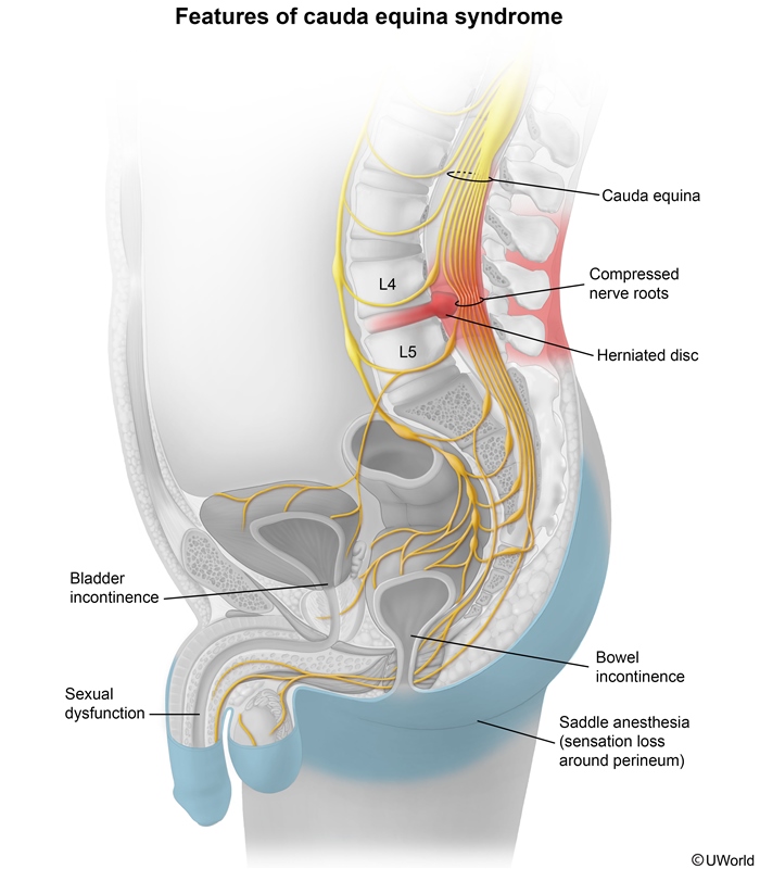
Cauda equina syndrome (CES) is a serious, often surgical, neurologic emergency characterized by compression of the cauda equina nerves in the lumbosacral spine. CES can lead to severe motor, sensory, and autonomic dysfunction, primarily affecting the lower limbs, bowel, and bladder.
Pathogenesis and risk factors
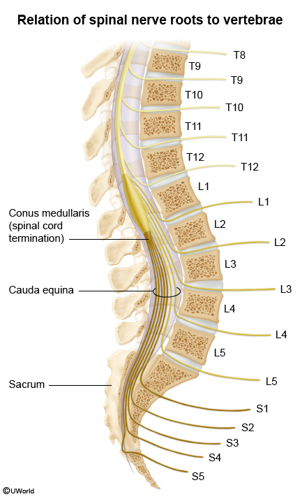
In an adult, the spinal cord terminates in a tapering fashion as the conus medullaris (T12-S4) at the L1-L2 vertebral level (Figure 1). The collection of spinal nerves below this point exits inferiorly through their respective intervertebral foramina and is referred to as the cauda equina (horse's tail). The cauda equina is composed of nerve roots for L2-L5, S1-S5, and the coccygeal nerve; these nerves are responsible for motor and sensory innervation to the lower limbs and pelvic organs, primarily via the sciatic nerve, pudendal nerve, and/or pelvic splanchnic nerves. The pelvic splanchnic nerves (S2-S4) provide parasympathetic innervation to the hindgut, bladder, and urinary sphincters that promotes peristalsis, bladder emptying, and pelvic floor relaxation during defecation.
Continue Learning with UWorld
Get the full Cauda Equina Syndrome article plus rich visuals, real-world cases, and in-depth insights from medical experts, all available through the UWorld Medical Library.
Unlock Full AccessFigures