Neuroanatomy: Spinal Cord
Article Sections
Introduction
The spinal cord is a vital structure in the central nervous system, extending from the medulla at the base of the brain to the level of the first or second lumbar vertebrae. It serves as a major conduit for transmitting neural signals between the brain and the rest of the body. Understanding the neuroanatomy of the spinal cord is crucial for localizing lesions and correlating specific clinical signs with different spinal cord regions and tracts.
Basic structure of the spinal cord
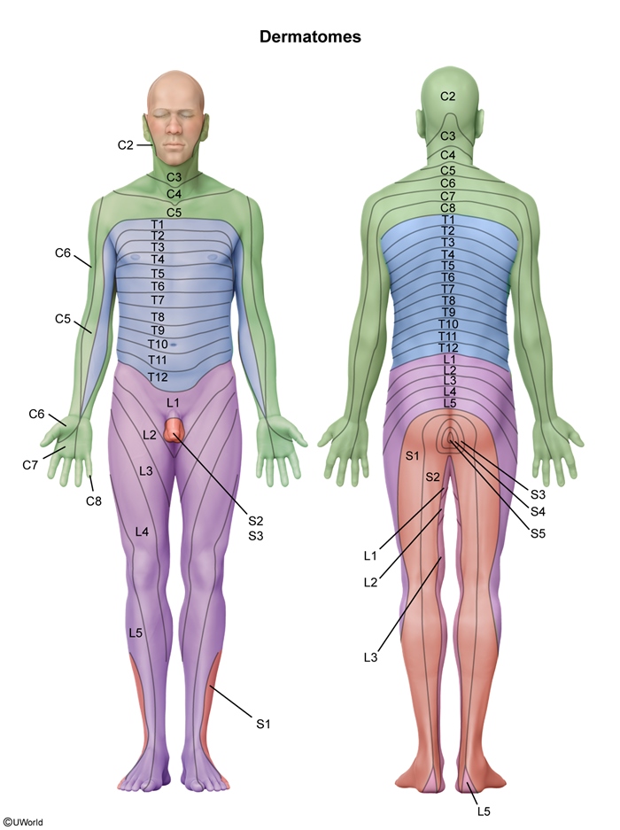
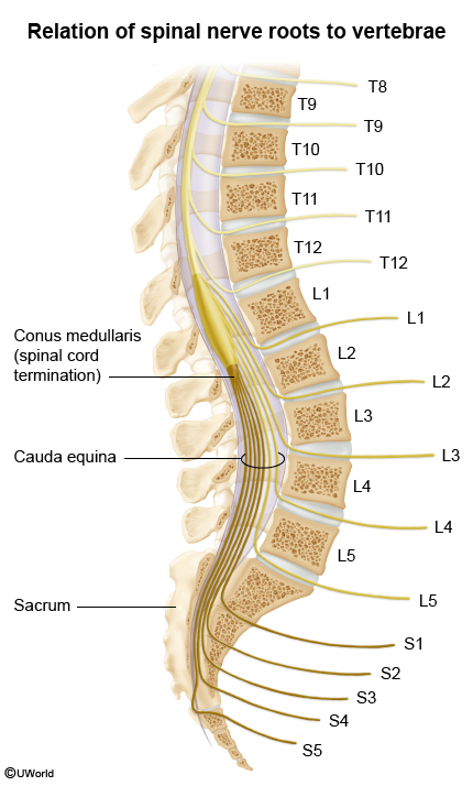
The spinal cord is organized into 31 segments (8 cervical, 12 thoracic, 5 lumbar, 5 sacral, and 1 coccygeal). Each segment of the spinal cord gives rise to sensory (afferent) and motor (efferent) nerve roots, each corresponding to pairs of spinal nerves that innervate specific myotomes and dermatomes (
Continue Learning with UWorld
Get the full Neuroanatomy: Spinal Cord article plus rich visuals, real-world cases, and in-depth insights from medical experts, all available through the UWorld Medical Library.
Unlock Full AccessFigures