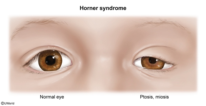
Horner Syndrome
Article Sections
Introduction
Horner syndrome is a neurologic disorder characterized by a classic triad of ptosis, miosis, and anhidrosis, resulting from disruption of the sympathetic nerve pathways supplying the eye and surrounding facial structures. The condition can arise from a variety of etiologies ranging from benign to life-threatening, making its identification and evaluation critical in clinical practice.
Anatomy and pathophysiology
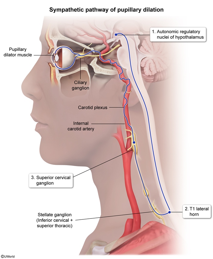
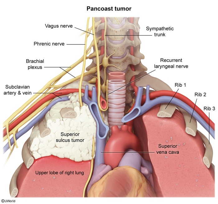
In response to dim light, input from the oculosympathetic pathway initiates pupillary dilation, allowing more light to reach the retina (Figure 1). The oculosympathetic innervation of the eye occurs via the following pathway:
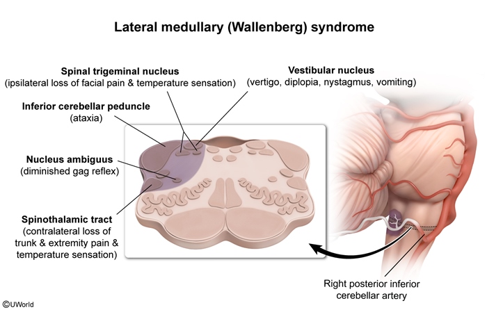
- First-order axons originate in the hypothalamus and descend in the lateral brainstem (next to the spinothalamic tract).
- They exit the spinal cord between C8 and T2 to reach the stellate ganglion in the paravertebral sympathetic chain.
Continue Learning with UWorld
Get the full Horner Syndrome article plus rich visuals, real-world cases, and in-depth insights from medical experts, all available through the UWorld Medical Library.
Unlock Full AccessFigures