Vitamin D Deficiency Rickets
Article Sections
Introduction
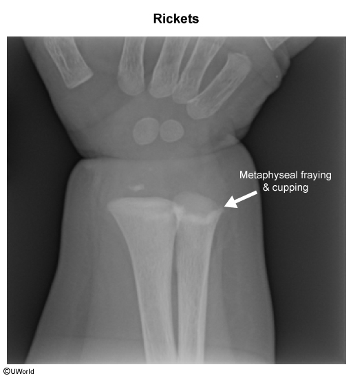
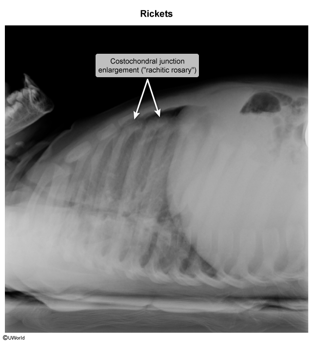
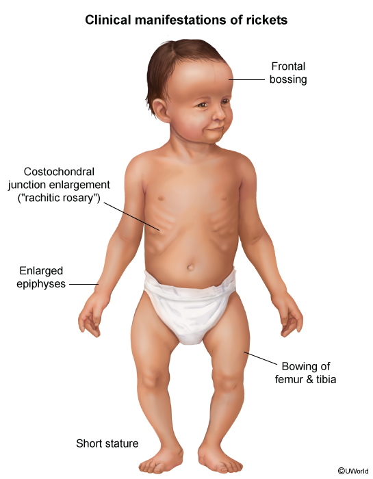
Vitamin D deficiency rickets is a preventable disease caused by decreased mineralization of growth plates and softening of bones during childhood. Patients classically present with skeletal changes, including widening of the wrists and bowing of the extremities.
Pathogenesis
Most bones, including long bones and vertebral bodies, develop through endochondral ossification, a process that begins during embryonic development. Endochondral ossification starts with mesenchymal cells differentiating into chondrocytes, forming a hyaline cartilage model for future bone. This cartilage template is then gradually replaced by bone through a series of steps. First, the cartilage matrix becomes mineralized by the deposition of calcium and phosphate into the osteoid (unmineralized bone matrix). Mineralization allows blood vessels to invade, triggering osteoblasts to create new bone matrices, converting the tissue to bone. Ossification begins in the central shaft of the bone (diaphysis) and advances toward the ends (epiphysis), where new cartilage continues to form. Ongoing ossification at these growth plates enables the replacement of cartilage with bone, supporting continuous linear growth throughout childhood and adolescence.
Continue Learning with UWorld
Get the full Vitamin D Deficiency Rickets article plus rich visuals, real-world cases, and in-depth insights from medical experts, all available through the UWorld Medical Library.
Unlock Full AccessFigures

Images