Embryology Of The Renal System
Article Sections
Introduction
Renal development is a complex, highly regulated process involving multiple embryologic structures that contribute to the formation of the kidneys, ureters, and bladder. A thorough understanding of renal embryology is essential for the diagnosis and management of congenital renal anomalies.
Overview
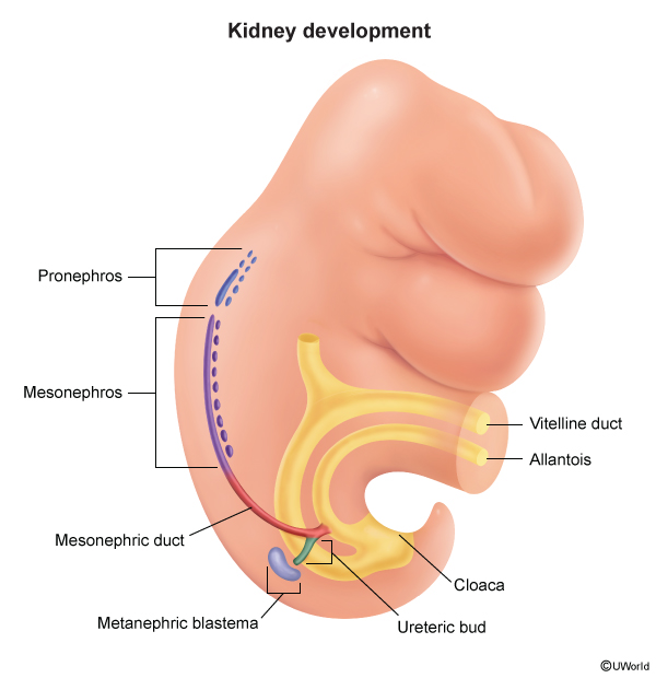
Renal development begins in early embryogenesis with the formation of the nephrogenic cord from intermediate mesoderm in the lower thoracic, lumbar, and sacral regions. Renal development occurs in a cranial-to-caudal sequence and progresses through 3 sequential stages (Figure 1), as follows:
- Pronephros (weeks 3-4): Derived from intermediate mesoderm in the cervical region. The pronephros is composed of 7-10 groups of nephrotomes, or rudimentary, nonfunctional excretory units that regress before the end of the fourth week of gestation. The pronephric ducts persist temporarily and initiate formation of the mesonephric duct.
Continue Learning with UWorld
Get the full Embryology Of The Renal System article plus rich visuals, real-world cases, and in-depth insights from medical experts, all available through the UWorld Medical Library.
Unlock Full AccessFigures