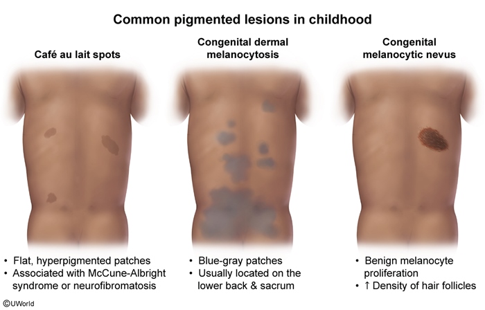
Congenital Melanocytic Nevi
Article Sections
Introduction
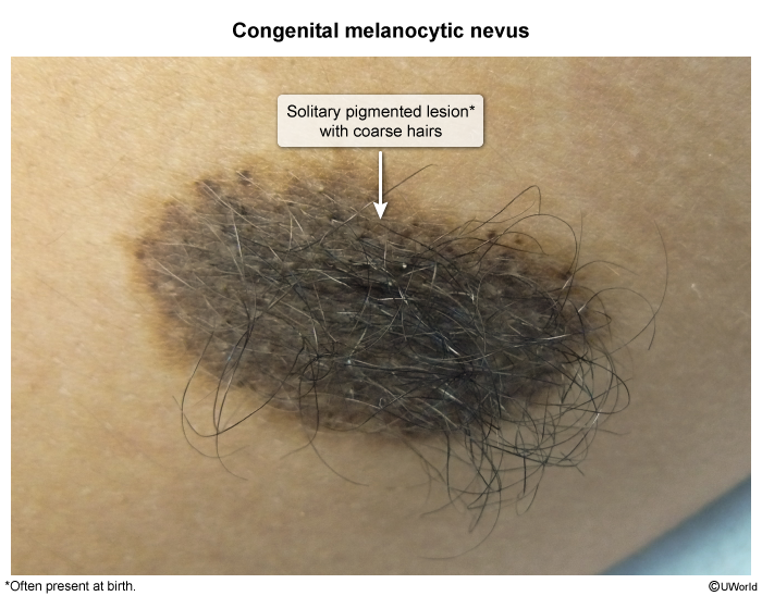
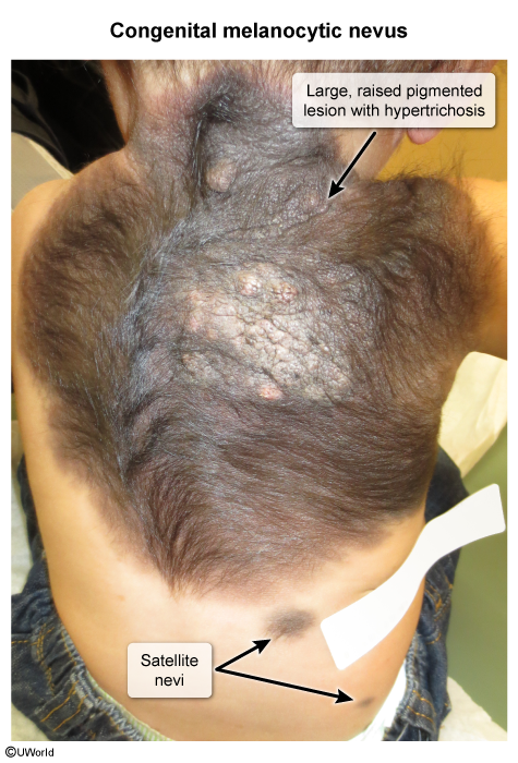
Congenital melanocytic nevi (CMN) are hyperpigmented lesions that are present at birth or appear within the first few months of life. These nevi are due to melanocyte proliferation and vary in size, with larger lesions carrying an increased risk for malignant transformation.
Pathogenesis
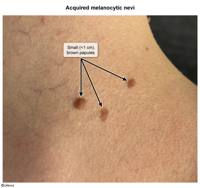
CMNs are caused by a benign proliferation of melanocyte cells. In contrast to the familial and ultraviolet-related pathogenesis of acquired nevi, CMNs likely arise from abnormal migration or proliferation of melanocytes as they migrate from the neural crest to the epidermis. Most cases are sporadic, but large CMNs are associated with somatic mutations in the NRAS gene during embryonic development.
Histologically, clusters of melanocytes in CMNs can extend from the epidermis to the dermis, subcutaneous fat, or muscle, with extension along hair follicles and sweat glands.
Continue Learning with UWorld
Get the full Congenital Melanocytic Nevi article plus rich visuals, real-world cases, and in-depth insights from medical experts, all available through the UWorld Medical Library.
Unlock Full AccessFigures
Images