Valve Disorders: Mitral Regurgitation
Article Sections
Introduction
Mitral regurgitation (MR) results from impaired closure of the mitral valve leaflets, leading to backflow of blood from the left ventricle into the left atrium during systole. It can be caused by structural abnormalities of the mitral valve apparatus itself (primary MR) or from left ventricular dysfunction that impairs valve closure despite a structurally normal valve (secondary or functional MR). Severe MR causes substantial hemodynamic disruption that leads to heart failure.
Pathophysiology
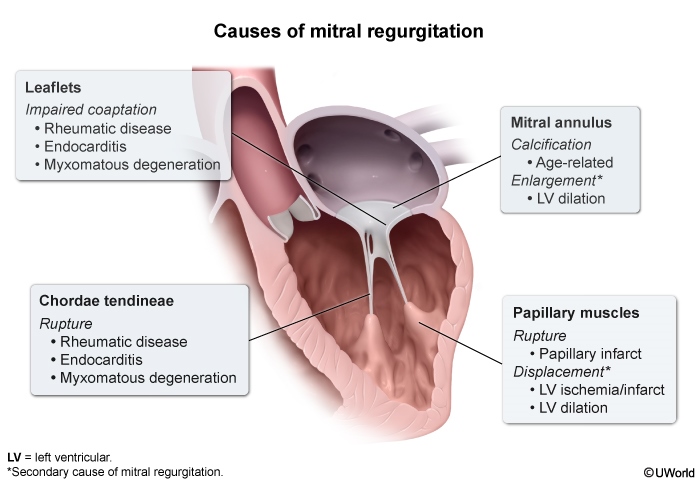
The mitral valve is composed of an anterior leaflet and a posterior leaflet, each supported by chordae tendineae attached to a papillary muscle. MR most commonly results from disruption of one or more of these 4 components (primary MR) (Figure 1). Less commonly, a separate disease process (eg, ischemic heart disease, dilated cardiomyopathy) causes left ventricular dysfunction that leads to impaired mitral valve closure despite normal valve structure (
Continue Learning with UWorld
Get the full Valve Disorders: Mitral Regurgitation article plus rich visuals, real-world cases, and in-depth insights from medical experts, all available through the UWorld Medical Library.
Unlock Full AccessFigures